
Inpatient Services with an Innovative Approach
For nearly 70 years, Advanced Cardiovascular Specialists has been delivering comprehensive, cutting-edge heart health care to patients in Southeast Texas. Our team of experienced cardiologists, cardiovascular disease specialists, and interventional cardiology experts lead the region in groundbreaking procedures, innovative therapies, and advanced diagnostics, and we’re committed to providing you with the highest level of care for achieving optimal heart health.
At Advanced Cardiovascular Specialists, our inpatient cardiovascular services include everything from advanced imaging to various interventions and therapies and can be performed right here in our Cardiovascular Diagnostic Center, a free-standing outpatient Cath Lab adjacent to our offices. We leverage the latest technology for the best in comprehensive, precision-guided procedures for the most accurate diagnosis and effective treatment planning.
Inpatient services at Advanced Cardiovascular Specialists include:
Balloon Angioplasty
Percutaneous Coronary Interventions This is a term for all of the procedures that we perform in the arteries that feed the heart with blood to treat narrowings, blockages or plaque with the use of catheter-based treatments. These interventions …

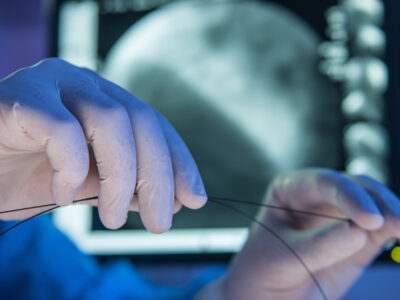
Cardiac Catheterization and Coronary Angiography
Coronary Angiography and Left Heart Catheterization are common terms for the same procedure that is also commonly referred to as the “Dye Test”. This procedure is the best test to define the exact location and severity of narrowings or blockages in …
Carotid and Peripheral Angiography
These tests are similar to Coronary Angiography. In this situation, the catheters’ tips are directed to different arteries. The risks of these procedures are similar to the risks listed for Cardiac Catheterization. In the case of Carotid and Cerebral …

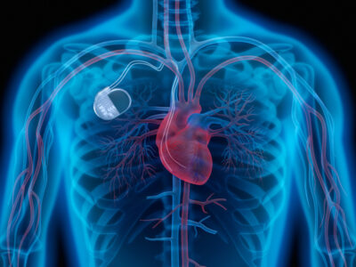
Implantable Cardiac Defibrillators
Implantable Cardiac Defibrillators are similar to pacemakers and the surgery required to place them is much the same. These are used for treatment of episodes of rapid heartbeat and/or fibrillation; especially of the lower chambers of the heart. They …
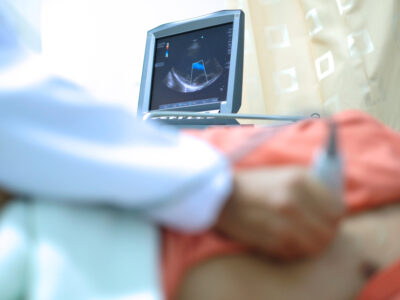
Intravascular Ultrasounds (IVUS)
Intravascular ultrasound (IVUS) is an advanced medical imaging procedure that uses sound waves to produce detailed images of the heart and blood vessels from within the body. This cutting-edge technology provides invaluable insights into the …
Pacemaker Implantation
Pacemakers are devices used to prevent the heart slowing excessively. If a patient has pauses or a slow heartbeat, which often leads to symptoms of fatigue, lack of energy or passing out, a pacemaker may be indicated. A small surgery is required to …

Rotablation (Rotational Atherectomy)
Rotablation, also known as rotational atherectomy (RA) or simply atherectomy, is a specialized medical procedure designed to remove plaque buildup and open blockages in coronary arteries. This technique is particularly useful in situations where …
Stent Placement to include Drug Eluding Stents
Percutaneous Coronary Interventions This is a term for all of the procedures that we perform in the arteries that feed the heart with blood to treat narrowings, blockages or plaque with the use of catheter-based treatments. These interventions …

Myocardial Perfusion Imaging (MPI)
Myocardial Perfusion Imaging (MPI) is a non-invasive imaging test that evaluates how well blood flows through your heart muscle. This advanced diagnostic tool can identify areas of the heart muscle that aren’t receiving adequate blood supply and …
Tilt Table Testing
Tilt Table Testing is an evaluation for the tendency to pass out under certain conditions. People who are especially prone to passing out may suffer from neurocardiogenic syncope. This syndrome is an abnormal exaggeration of a normal reflex and can …
Transesophageal Echocardiography
Transesophageal Echocardiogaphy is a specialized form of ultrasound where the probe is at the tip of a long, thin tube. A patient is given sedation and this tube is placed through the mouth and into the esophagus and stomach in order to make …
More about Inpatient Services at Advanced Cardiovascular Specialists:
Percutaneous Coronary Interventions, which include all of the procedures performed in the arteries that feed the heart with blood. These procedures use catheter-based treatments to treat narrowings, blockages, and/or plaque and prop the artery open for better blood flow.
Balloon Angioplasty, a minimally-invasive procedure that uses a balloon to open or widen a blocked coronary artery.
Cutting Balloons, a specific type of angioplasty device used when a traditional balloon is not effective, treating coronary artery conditions by incising and dilating the affected vessel.
Rotablation (Rotational Atherectomy), a specialized medical procedure designed to remove plaque buildup and open blockages in coronary arteries, especially where traditional angioplasty may not be effective.
Brachytherapy, a procedure that uses radiation to remove scar tissue that can build up around stents and prevent opened arteries from narrowing again, especially for patients with multiple stents or recurring scar tissue development (in-stent restenosis).
Stent Placement to include Drug Eluting Stents (DES). Stent placement typically follows angioplasty, and involves implanting a small metal mesh tube device in the newly-opened artery wall where it acts as a scaffold to keep the artery open. DES can be more effective at preventing recurring blockages.
Cardiac Catheterization and Coronary Angiography. Also referred to as the “Dye Test,” this procedure is the gold standard for defining the exact location and severity of narrowings or blockages in the coronary arteries. It is necessary before performing any coronary intervention and provides valuable information about the valves and ventricular function on the left side of the heart. Other cardiac catheterization procedures include:
Intravascular Ultrasounds (IVUS), an advanced medical imaging procedure that uses sound waves to produce detailed images of the heart and blood vessels.
Carotid, Peripheral, and Cerebral Angiography, diagnostic procedures that use x-rays and a contrast material to examine the veins and arteries that supply blood to the brain (carotid and cerebral) and the lower extremities (peripheral) to identify blockages, narrowings, or abnormalities.
Implantable Cardiac Defibrillators. Similar to pacemakers, Implantable Cardiac Defibrillators are used to treat episodes of rapid heartbeat and/or fibrillation, pacing or shocking the heart internally to prevent episodes of sudden cardiac death caused by arrhythmia.
Pacemaker Implantation, in which a small electronic device is placed just below the collarbone to regulate electrical problems with the heart and prevent the heart from slowing excessively.
Myocardial Perfusion Imaging (MPI), or Nuclear Stress Testing, a non-invasive imaging test that evaluates how well blood flows through your heart muscle. It can identify areas of the heart that aren’t receiving adequate blood supply and assess the heart muscle’s pumping efficiency.
Tilt Table Testing is an evaluation for patients with a tendency to pass out, faint, or become dizzy under certain conditions. The Tilt Table simulates standing up to recreate the effect on your blood pressure and heart rate at various angles.
Transesophageal Echocardiography, a specialized form of ultrasound where the probe is at the tip of a long, thin tube, bypassing bone and lung tissue and providing more detailed pictures of the heart valves.
Industry-Leading Inpatient Care at Advanced Cardiovascular Specialists
At Advanced Cardiovascular Specialists, our experienced team of cardiologists and technicians is dedicated to delivering the highest quality of care with the highest level of compassion. Your comfort and confidence are prioritized every step of the way, which means our specialists share their extensive experience in both implementation and results interpretation with you to ensure you are informed and any concerns are addressed. Then, your physician will work with you to develop a personalized treatment plan that has only one goal—the best outcome for your heart health.
For the team of multidisciplinary experts at Advanced Cardiovascular Specialists, caring for cardiovascular health is about more than medicine; it’s about caring for the whole patient with honesty, integrity, and dignity. With five physicians across four locations, our physicians can provide innovative, leading-edge inpatient services when and where our patients need them most. Schedule an appointment today and experience the difference exceptional cardiovascular care can make for your heart’s health.