
Outpatient Services Delivered with Outstanding Expertise
For nearly 70 years, Advanced Cardiovascular Specialists has been a trusted leader in heart health care for Southeast Texas. Our team of expert cardiologists, cardiovascular disease specialists, and interventional cardiologists is dedicated to advancing heart care through innovative therapies, groundbreaking procedures, and state-of-the-art diagnostics. We are committed to providing every patient with personalized, high-quality care to support their journey to optimal heart health.
Our outpatient cardiovascular services offer a seamless blend of cardiovascular exams, advanced imaging services, and diagnostic testing—all delivered at our Cardiovascular Diagnostic Center. Conveniently located adjacent to our offices, this free-standing outpatient Cath Lab is equipped with the latest technology to ensure precise diagnoses and tailored treatment plans. At Advanced Cardiovascular Specialists, we combine expertise, innovation, and compassionate care to deliver exceptional outcomes for every patient.
Outpatient services at Advanced Cardiovascular Specialists include:
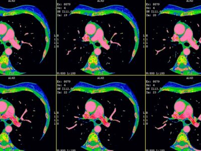
Cardiac CT Scan
64 Slice Cardiac Computed Tomography Scan This marvel of modern medicine images your body’s vital organs and tissues. It captures your health from the inside out, revealing build-up in your coronary arteries, nodules in your lungs, and potential …
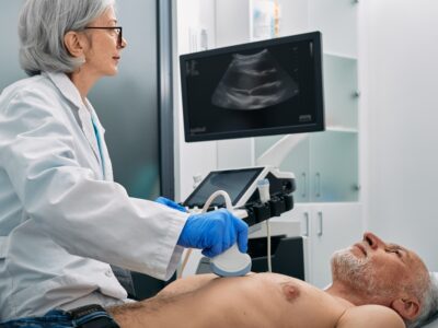
Carotid Ultrasounds
Vascular Ultrasound uses the same technology as Echo where sound waves are used to visualize the larger arteries such as the carotids in the neck, the aorta in the abdomen, the arteries leading to the kidneys, and the arteries leading to both legs. …
Coronary Artery Calcium Scoring (CACS)
Over half the people who died suddenly of a heart attack had no prior symptoms. To combat this, you can undergo a coronary artery calcium scoring (CACS) exam to scan the heart and detect calcium deposits along the walls of arteries. The test then …
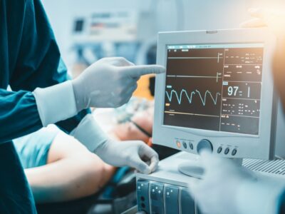
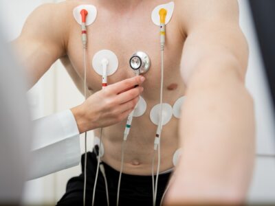
Electrocardiograms (ECGs or EKGs)
An electrocardiogram, commonly referred to as ECG or EKG, is a fundamental diagnostic tool in electrophysiology. This non-invasive procedure measures the electrical signals that regulate your heart’s activity. How Does an ECG Work? To conduct …
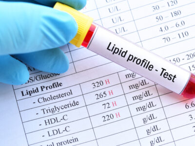
Lipid Center
Certain frequently performed lab tests can be obtained in our office with the convenience of a finger stick. For patients on chronic coumadin therapy, protimes can be measured with this test and results available while the patient waits (less than 5 …
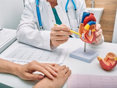
Nuclear Cardiology
Nuclear medicine provides functional imaging of the heart. It involves injecting medically safe radioisotopes to detect the presence of heart disease. Stress myocardial perfusion imaging adds significant diagnostic and prognostic information …

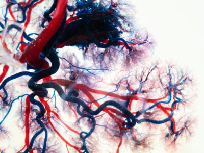
Peripheral Vascular Studies
Vascular Ultrasound uses the same technology as Echo where sound waves are used to visualize the larger arteries such as the carotids in the neck, the aorta in the abdomen, the arteries leading to the kidneys, and the arteries leading to both legs. …
Protime Clinic
Certain frequently performed lab tests can be obtained in our office with the convenience of a finger stick. For patients on chronic coumadin therapy, protimes can be measured with this test and results available while the patient waits (less than 5 …
Resting and Stress Echocardiograms
Resting and Stress Echo is an ultrasound of the heart at rest and is then repeated at peak exercise and allows us to see the movement of the walls of the pumping chamber of the heart and compare them at rest and with exercise. Echocardiography is an …
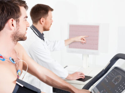
Treadmill Testing
Basic treadmill exercise testing involves EKG monitoring at rest and continuously while walking on a treadmill, which starts at a slow rate and with a mild incline, increases every three minutes until a person has reached their target heart rate or …
More about Outpatient Services at Advanced Cardiovascular Specialists:
Cardiac CT Scan. The 64-Slice Cardiac Computed Tomography Scan captures your health from the inside out, revealing build-up in the coronary arteries, nodules in the lungs, and potential tumors in the abdomen and pelvis. As a non-invasive angiography–essentially a “dye test” without the catheter–the Cardiac CT Scan can detect the smallest amount of plaque even if no calcium is present.
Carotid/Vascular Ultrasounds, a non-invasive technology that uses sound waves to visualize plaque, narrowing, or blockages in the larger arteries such as the carotids in the neck, the aorta in the abdomen, the arteries leading to the kidneys, and the arteries leading to both legs (peripheral vascular studies).
Coronary Artery Calcium Scoring (CACS), an exam that scans the heart, detects calcium deposits along the walls of the arteries, and immediately produces a calcium score that identifies your level of deposits and can help measure your potential for heart disease.
Electrocardiograms (ECGs or EKGs), a diagnostic tool that measures the electrical signals regulating your heart’s activity and provides valuable insights into various aspects of your heart’s health. Advanced Cardiovascular Specialists uses 12-lead ECGs, which provide the most advanced, comprehensive view of your heart’s electrical activity from multiple angles.
Nuclear Cardiology, which provides functional imaging of the heart by injecting medically safe radioisotopes to detect the presence of heart disease. Nuclear medicine services include Cardiac PET, Myocardial perfusion studies, perfusion imaging, pharmacological stress tests, and ventricular function.
Peripheral Vascular Studies, non-invasive tests used to evaluate blood flow in the arteries and veins of the legs and feet and diagnose or monitor conditions like peripheral artery disease (PAD), deep vein thrombosis (DVT), and varicose veins.
Protime and Lipid Clinic, where we can perform a variety of routine lab tests with the convenience of a finger stick and immediate results. Cholesterol and liver function tests can be performed here, as well as protimes for patients on chronic coumadin therapy, allowing for more timely adjustments without having to wait for or schedule a follow-up.
Resting and Stress Echocardiograms, ultrasound examinations of the heart at rest and at peak exercise that allow your physician to evaluate the movement of the walls of the pumping chamber of the heart, accurately measure the heart size, look for fluid in the sack surrounding the heart, evaluate blood flow through the valves, and evaluate overall heart muscle function.
Treadmill Testing, which involves EKG monitoring at rest and continuously while walking on a treadmill to highlight any blockages or narrowing of the coronary arteries.
Leading-Edge Outpatient Care at Advanced Cardiovascular Specialists
At Advanced Cardiovascular Specialists, our team of dedicated cardiologists and technicians combines medical expertise with genuine compassion to deliver exceptional care. We prioritize your comfort and confidence at every step, ensuring you are fully informed about your diagnosis and treatment options. By providing clear explanations and addressing your concerns, we empower you to make confident decisions about your heart health. From there, your physician will collaborate with you to create a personalized treatment plan, all with one goal in mind—achieving the best possible outcome for your heart.
For the multidisciplinary team at Advanced Cardiovascular Specialists, cardiovascular care goes beyond medicine—it’s about treating the whole person with honesty, integrity, and respect. With five skilled physicians across four convenient locations, we’re proud to offer cutting-edge inpatient services tailored to meet your needs. Let us support your heart health journey with expertise, compassion, and personalized care. Schedule your appointment today and discover the difference that truly patient-focused cardiovascular care can make.